Epithelial Tissue: Skin, Barriers, and Wound Healing in Physical Therapy
Learn how epithelial tissue protects the body, how skin wounds heal, and how physical therapy supports skin integrity and wound management.
When most people think of physical therapy, they picture exercises for joint injuries or stroke rehabilitation. But physical therapists also play a crucial role in wound care, skin integrity, and the management of conditions where the body’s first line of defense — epithelial tissue — has been compromised.
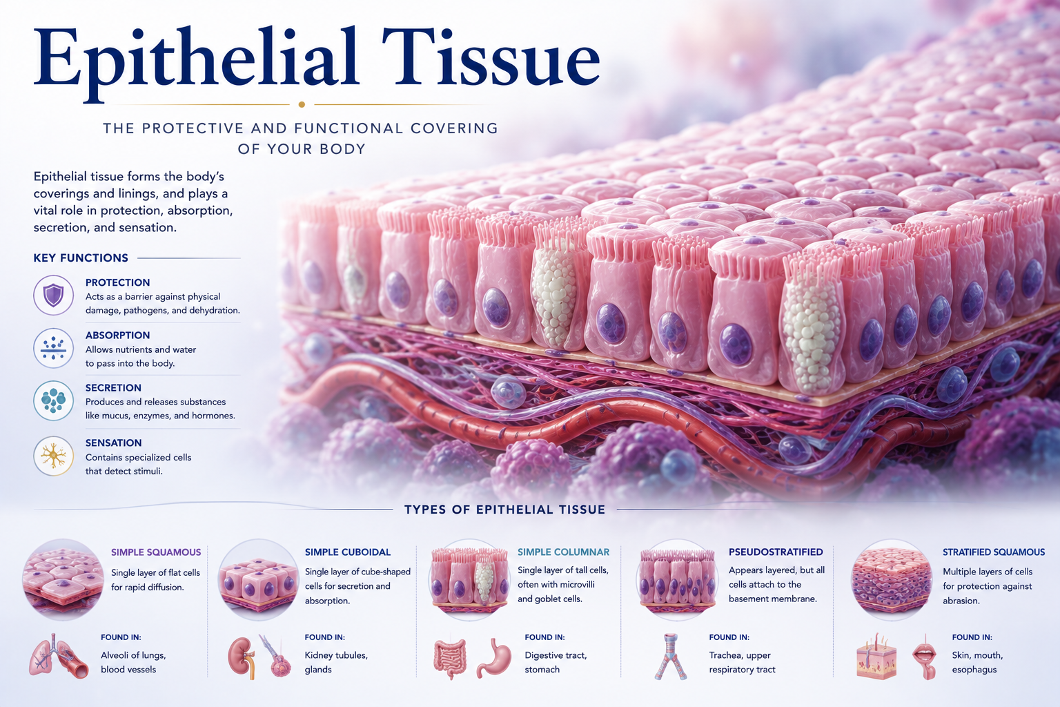
Epithelial tissue is the tissue that lines and covers. It forms the skin on the outside of the body and lines the inner surfaces of organs and vessels. Understanding how it is structured and how it heals is essential for physical therapists working with patients who have pressure ulcers, post-surgical wounds, burns, amputations, or conditions that impair skin integrity.
What Is Epithelial Tissue?
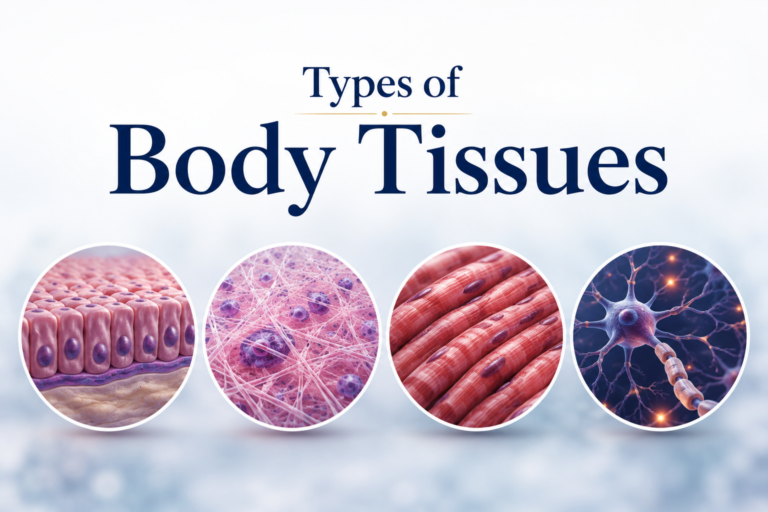
Epithelial tissue is characterized by cells that are tightly packed together, covering surfaces and forming barriers. Unlike connective tissue — which has cells scattered within an extracellular matrix — epithelial cells form continuous sheets with minimal space between them, connected by specialized cell junctions.
Epithelial tissue is always positioned at a boundary: between the body and the outside environment, or between different internal compartments. It receives no direct blood supply (it is avascular) but is nourished by diffusion from underlying connective tissue.
Epithelial tissue is classified by two criteria:
- Number of layers: Simple (single layer) or stratified (multiple layers).
- Cell shape: Squamous (flat), cuboidal, or columnar.
The most complex epithelium in the body is the stratified squamous epithelium of the skin — multiple layers of cells that provide the primary physical barrier between the body and the external world.
The Skin: Structure and Function
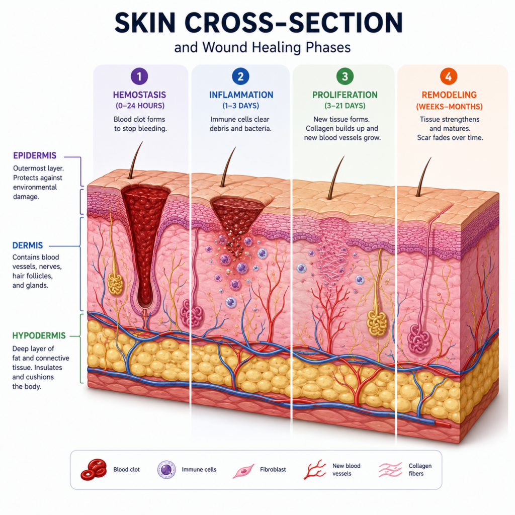
The skin is the largest organ in the human body, covering approximately 2 square meters in an average adult. It is composed of two main layers:
The epidermis is the outermost layer, composed of stratified squamous epithelial cells called keratinocytes. The deepest layer (stratum basale) contains actively dividing cells that constantly regenerate the epithelium. As these cells divide, older cells are pushed toward the surface, where they flatten, accumulate the protein keratin, and eventually die and are shed. This continuous renewal process takes approximately 28-35 days under normal conditions.
Embedded within the epidermis are specialized cell types including melanocytes (which produce the pigment melanin, providing UV protection) and Langerhans cells (immune cells that detect pathogens at the skin surface).
The dermis is the deeper layer of the skin, composed of dense connective tissue rich in collagen and elastin fibers. It contains blood vessels, lymphatic vessels, nerves, hair follicles, sweat glands, and sebaceous (oil) glands. The dermis provides the skin’s structural strength and elasticity.
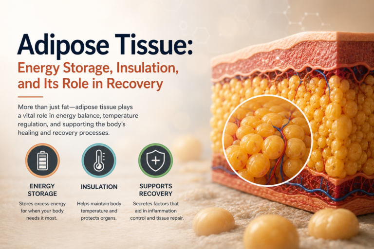
Beneath the dermis lies the hypodermis (subcutaneous tissue) — a layer of loose connective tissue and adipose (fat) tissue that provides insulation, cushioning, and energy storage.
Functions of Epithelial Tissue in the Body
Skin and epithelial tissue perform several functions critical to health:
Protection: The epidermis acts as a physical barrier against mechanical injury, pathogens, UV radiation, and water loss. Keratin makes it tough and waterproof. Tight junctions between cells prevent pathogens from penetrating.
Sensation: The dermis contains a rich network of sensory nerve endings that detect touch, pressure, vibration, temperature, and pain — providing essential information for motor control and proprioception.
Thermoregulation: Sweat glands in the dermis produce sweat that evaporates and cools the body. Blood vessels in the dermis dilate or constrict to increase or decrease heat loss from the skin surface.
Vitamin D synthesis: Exposure to UV radiation converts precursor molecules in the skin into vitamin D, which is essential for calcium absorption and bone health — directly relevant to physical therapy patients managing osteoporosis and fractures.
Immune defense: Langerhans cells in the epidermis detect and respond to pathogens. The skin’s acidic pH and antimicrobial peptides provide additional protection against infection.
Wound Healing: A Staged Biological Process
When skin is breached — by injury, surgery, or pressure — the body initiates a complex, well-orchestrated repair process. Understanding the stages of wound healing helps physical therapists time their interventions appropriately and avoid inadvertently disrupting the healing process.
Stage 1 — Hemostasis (immediate): Bleeding is controlled by platelet aggregation and clot formation. Platelets also release growth factors that initiate the healing cascade.
Stage 2 — Inflammation (days 1-5): White blood cells infiltrate the wound, cleaning debris and bacteria. The wound appears red, swollen, and warm. This stage is essential — it sets up the environment for repair — but excessive or prolonged inflammation impairs healing.
Stage 3 — Proliferation (days 5-21): Fibroblasts migrate into the wound and produce collagen, filling the defect with granulation tissue. Epithelial cells at the wound edges begin dividing and migrating across the granulation tissue to close the wound — a process called re-epithelialization. New blood vessels grow into the wound (angiogenesis).
Stage 4 — Remodeling (weeks to years): The initial scar tissue — which is weaker and less organized than normal skin — is gradually remodeled. Type III collagen is replaced by stronger Type I collagen. The scar matures, typically flattening, lightening, and gaining strength over months to years. Even a well-healed scar only reaches approximately 70-80% of the tensile strength of uninjured skin.
Physical Therapy in Wound Care
Physical therapists contribute significantly to wound care and management of skin integrity:
Pressure ulcer prevention involves repositioning protocols, positioning equipment, and therapeutic exercise to maintain mobility and reduce sustained pressure over bony prominences. Exercise improves local circulation, enhancing tissue perfusion and resilience.
Post-surgical wound management includes education about wound care, preventing complications, managing scar tissue, and facilitating functional recovery. Scar management — including massage, silicone gel, and active stretching — helps prevent restrictive scar formation that limits joint movement.
Burns rehabilitation is a specialized area of physical therapy involving range of motion exercises, splinting, compression garment use, and functional retraining. Preventing contractures (shortening of scar tissue across joints) is a primary goal.
Lymphedema management addresses chronic swelling caused by compromised lymphatic drainage — often seen after cancer surgery — through manual lymphatic drainage, compression therapy, and exercise.
Epithelial Lining of Internal Structures
Beyond skin, epithelial tissue lines the lungs, gastrointestinal tract, urinary tract, and reproductive organs. Physical therapy intersects with these systems in specialized ways — for example, pelvic floor physical therapy addresses conditions affecting the epithelial lining of the pelvic organs, and respiratory physical therapy addresses airway clearance in conditions where the respiratory epithelium is compromised.
Conclusion
Epithelial tissue is the body’s protective shield — a dynamic, self-renewing barrier that guards against physical damage, infection, and environmental hazards. When it is disrupted, the body launches a sophisticated repair process that physical therapy can support, guide, and optimize.
For physical therapists, understanding skin structure and wound healing biology enhances clinical decision-making — from timing the introduction of exercise to managing scar tissue to preventing complications in vulnerable patients. For patients, understanding these processes builds confidence in the healing journey and highlights the importance of consistent, appropriate physical therapy care.
References
- Ross, M.H., & Pawlina, W. (2020). Histology: A Text and Atlas (8th ed.). Wolters Kluwer.
- Mescher, A.L. (2021). Junqueira’s Basic Histology: Text and Atlas (16th ed.). McGraw-Hill.
Disclaimer: This article is for educational purposes only and does not constitute medical advice. Always consult a qualified healthcare professional for personal health concerns.
The site is written and curated by Paul Morgan, a graduate in Physiotherapy (2026), with a particular interest in cardiorespiratory and musculoskeletal physical therapy. Every article on this site is grounded in academic physiology and physical therapy coursework. Content reviewed for clinical accuracy before publishing.