Bone Tissue: Structure, Remodeling, and Why It Matters in Physical Therapy
Bone often gets treated as the fixed scaffolding the rest of physiology happens on — the thing that stays still while muscles, blood, and nerves do the interesting work. That’s a mistake. Bone is a living, constantly remodeling connective tissue, and understanding how it rebuilds itself is central to managing fractures, osteoporosis, and almost anything involving load-bearing rehabilitation.
What Bone Tissue Is Made Of
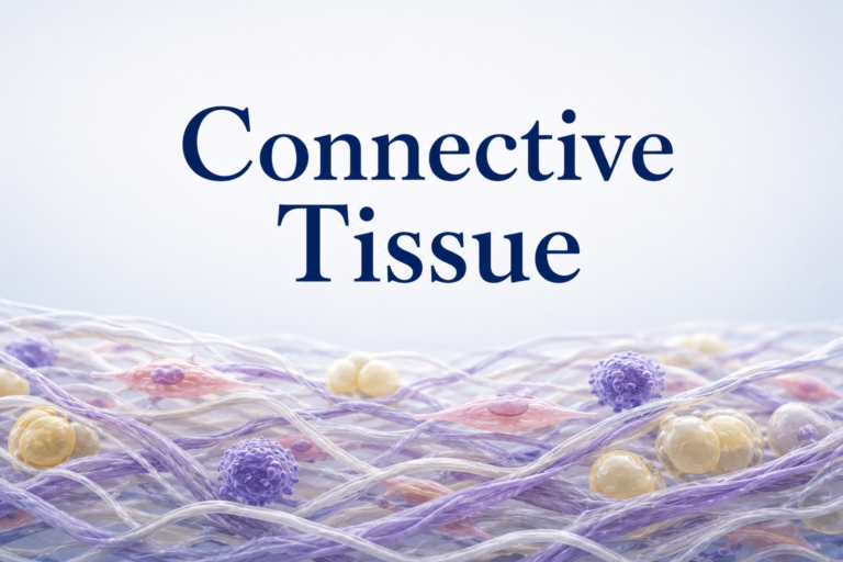
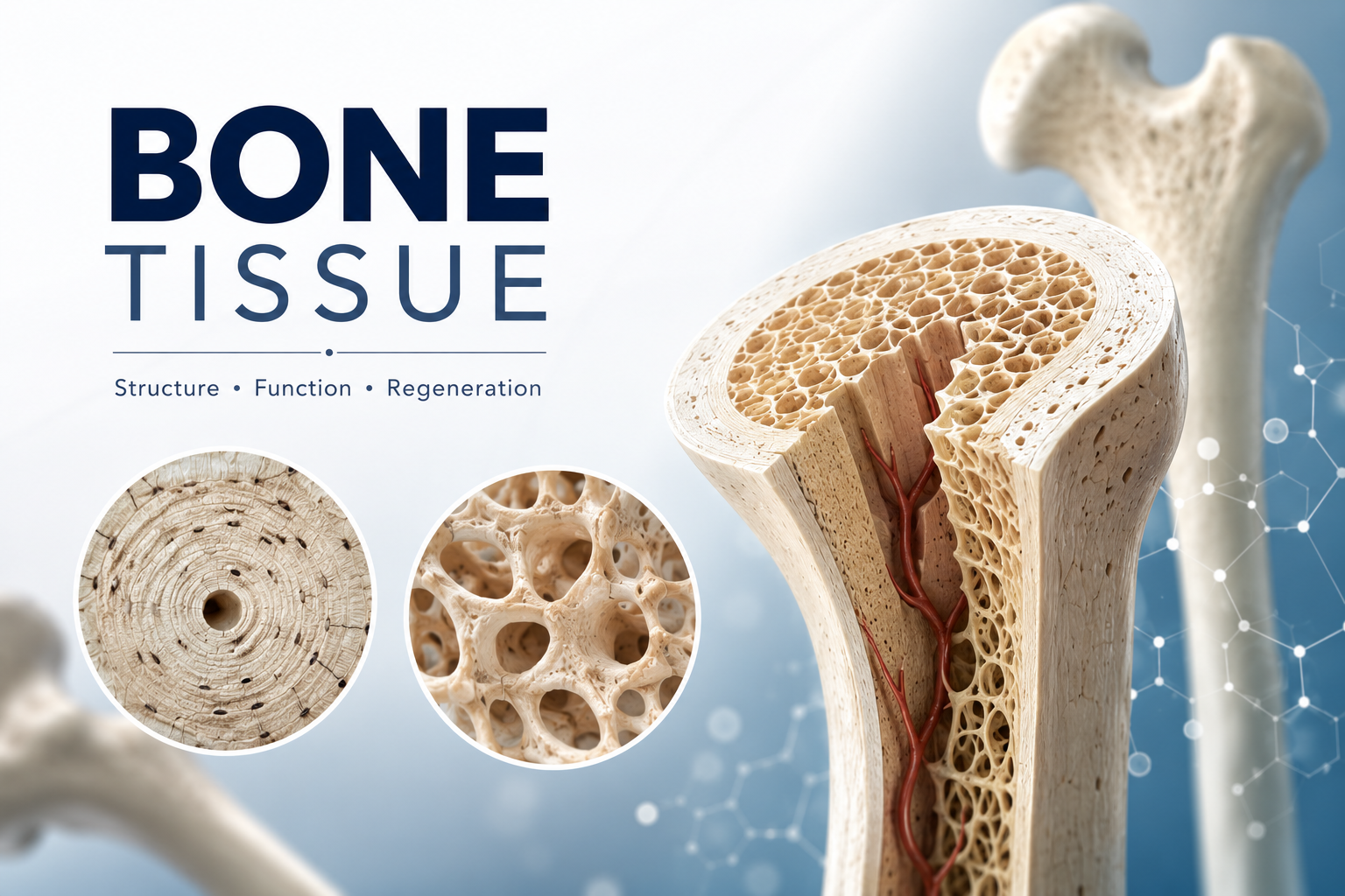
Bone is a connective tissue, which means it follows the same basic recipe as cartilage or blood: cells embedded in an extracellular matrix that determines the tissue’s properties. What makes bone distinct is that its matrix is mineralized. Roughly a third of that matrix is organic — mostly type I collagen, which gives bone tensile strength and a degree of flexibility — and the rest is inorganic, dominated by hydroxyapatite crystals (a calcium-phosphate compound) that give bone its compressive strength and rigidity. Take away the mineral and bone becomes rubbery; take away the collagen and it becomes brittle. The combination is what allows a femur to handle compressive load and the occasional sudden torque without snapping.
Three Cells, Three Jobs
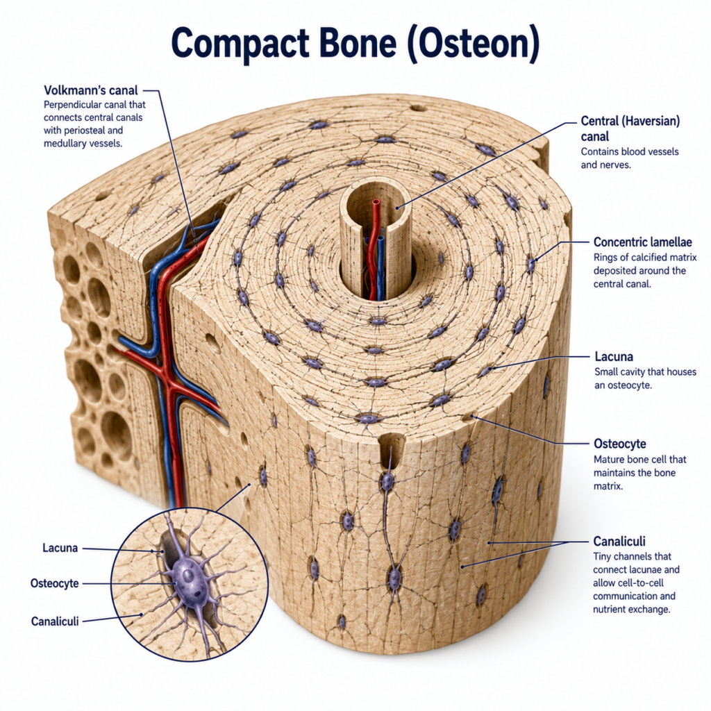
Bone tissue function comes down to three cell types working in a coordinated cycle. Osteoblasts synthesize new matrix — they are the construction crew, laying down collagen and triggering its mineralization. Once an osteoblast becomes fully surrounded by the matrix it built, it transitions into an osteocyte, a mature bone cell that no longer builds new tissue but maintains the existing matrix and communicates with neighboring cells through long cytoplasmic extensions running through tiny channels called canaliculi. Osteoclasts do the opposite job: they are large, multinucleated cells that resorb bone, dissolving the mineral matrix and releasing calcium and phosphate back into circulation.
This isn’t a one-time construction process — it’s continuous. Healthy bone replaces a meaningful percentage of its tissue every year through a cycle of resorption and formation, and the balance between osteoblast and osteoclast activity is what determines whether bone mass increases, holds steady, or declines.
Wolff’s Law: Why Loading Matters
In the late 1800s, the German anatomist Julius Wolff observed that bone adapts its internal architecture to the mechanical loads placed on it — a principle that still carries his name. Mechanical stress on bone activates osteocytes, which sense strain through fluid movement in the canalicular network and signal osteoblasts to reinforce loaded regions. Remove the load — through prolonged bed rest, immobilization, or spaceflight — and the opposite happens: osteoclast activity outpaces osteoblast activity, and bone density drops.
This is precisely why physical therapy matters so much after a fracture or during osteoporosis management. Weight-bearing exercise and progressive resistance loading aren’t just about muscle — they are a direct mechanical signal telling bone tissue to maintain or rebuild its density. A patient kept immobile longer than necessary doesn’t just lose muscle mass; they lose bone mass that can take far longer to recover.
Compact and Spongy Bone
Bone tissue comes in two architectural forms. Compact (cortical) bone is dense and forms the outer shell of most bones, organized into repeating cylindrical units called osteons, each built around a central Haversian canal that carries blood vessels and nerves. Spongy (cancellous) bone is found at the ends of long bones and inside flat bones, built from a lattice of trabeculae that’s lighter and less dense, but arranged along the lines of greatest mechanical stress — itself a demonstration of Wolff’s Law in action. Most bones combine both: a compact outer shell for strength, a spongy interior for weight savings and shock absorption.
Clinical Relevance: Fractures and Osteoporosis
Fracture healing essentially recapitulates parts of normal bone development. A hematoma forms first, followed by a soft callus of cartilage and woven bone, which is gradually remodeled into mature lamellar bone over weeks to months — a timeline that explains why premature loading can disrupt healing, while appropriately progressive loading later in the process accelerates it.
Osteoporosis, by contrast, is what happens when osteoclast activity chronically outpaces osteoblast activity, often driven by estrogen decline after menopause, vitamin D deficiency, or prolonged inactivity. The result is reduced bone density and a loss of trabecular architecture, raising fracture risk dramatically. This is one of the better-documented cases where physical therapy is not just supportive care but a primary intervention: targeted resistance and impact loading is one of the few interventions shown to meaningfully slow bone loss.
Conclusion
Bone is not inert scaffolding — it’s an actively managed tissue that responds, sometimes within weeks, to the mechanical demands placed on it. For a physical therapist, that means every loading decision in a rehabilitation program is also a conversation with osteoblasts and osteoclasts, whether the patient realizes it or not.
References
- Ross, M.H., & Pawlina, W. (2020). Histology: A Text and Atlas (8th ed.). Wolters Kluwer.
- Hall, J.E., & Hall, M.E. (2020). Guyton and Hall Textbook of Medical Physiology (14th ed.). Elsevier.
Further Reading
Disclaimer: This article is for educational purposes only and does not constitute medical advice. Always consult a qualified healthcare professional for personal health concerns.