Adipose Tissue: Energy Storage, Insulation, and Its Role in Recovery
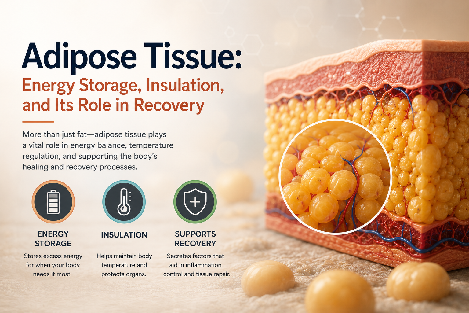
Adipose tissue has a branding problem. In casual conversation “fat” is shorthand for something to minimize, but as a tissue it performs jobs that have nothing to do with aesthetics: insulation, cushioning, hormone production, and one of the body’s two ways of generating heat. Understanding what adipose tissue actually does changes how it shows up in a rehabilitation context — not as a problem to solve, but as a tissue with real physiological functions that interact with healing, mobility, and metabolic health.
Adipocytes: Built to Store, But Not Just That
The defining cell of adipose tissue is the adipocyte, and most of what’s relevant about it depends on which of the two main types it is.
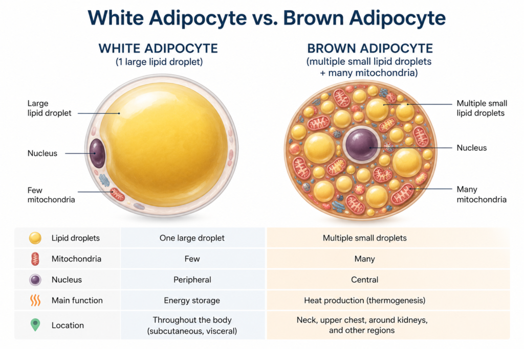
White adipocytes are built around a single, large lipid droplet that can occupy more than 90 percent of the cell’s volume, pushing the nucleus and other organelles to the periphery. This is the dominant fat type in adults and the body’s primary long-term energy reserve, but it does more than store triglycerides. White adipose tissue is also an active endocrine organ, secreting hormones — collectively called adipokines — including leptin, which signals satiety to the hypothalamus, and adiponectin, which influences insulin sensitivity. Chronic excess white adipose tissue, particularly visceral fat around abdominal organs, shifts this secretion pattern toward a pro-inflammatory profile, which is part of why obesity is linked to systemic low-grade inflammation that can slow tissue healing.
Brown adipocytes look structurally different: instead of one large droplet, they’re packed with multiple smaller lipid droplets and a strikingly high number of mitochondria, which is what gives the tissue its brown color. Those mitochondria contain a protein called UCP1 (uncoupling protein 1) that allows them to generate heat directly from stored energy rather than producing ATP — a process called non-shivering thermogenesis. Brown fat is abundant in infants, who rely on it heavily for temperature regulation, and persists in smaller, metabolically active deposits in adults, mainly around the neck and upper back.
Where Adipose Tissue Sits in the Body
Adipose tissue is distributed in functionally distinct depots. Subcutaneous fat sits beneath the skin and is the main contributor to thermal insulation and the body’s overall energy reserve. Visceral fat surrounds internal organs in the abdominal cavity and behaves quite differently — metabolically more active, more closely linked to insulin resistance, and the depot most responsible for the cardiometabolic risk associated with central obesity. A smaller but clinically relevant depot is the fat found within and around joints, bone marrow, and muscle (intramuscular adipose tissue), which becomes particularly relevant in conditions like sarcopenic obesity and prolonged disuse, where muscle tissue is partially replaced by fat infiltration.
Adipose Tissue and Mechanical Function
Beyond energy and hormones, adipose tissue plays a structural role that’s easy to overlook. Fat pads cushion joints — the infrapatellar fat pad behind the kneecap and the heel fat pad under the calcaneus are two examples directly relevant to orthopedic physical therapy, since both can become a source of localized pain when irritated or atrophied. Adipose tissue surrounding peripheral nerves and within fascial planes also contributes to gliding mechanics between tissue layers, which is part of the rationale behind some manual therapy and myofascial techniques.
Clinical Relevance for Rehabilitation
Body composition — the ratio of lean mass to fat mass, and where that fat is distributed — has measurable effects on rehabilitation outcomes. Excess visceral adiposity is associated with elevated systemic inflammatory markers, which can blunt the resolution phase of healing after injury or surgery. On the other end, very low fat mass, as seen in some eating disorders or overtraining, removes mechanical cushioning and can disrupt the hormonal signaling (including leptin) that supports normal bone density and reproductive physiology. Neither extreme is desirable, and physical therapists working with exercise prescription, particularly in metabolic or post-bariatric populations, are often implicitly managing adipose tissue health alongside muscle and connective tissue.
A related scenario worth recognizing is cachexia — severe, disease-driven fat and muscle wasting seen in some cancers, advanced heart failure, and chronic kidney disease. Unlike simple weight loss from reduced intake, cachexia is driven by systemic inflammatory signaling that actively breaks down both adipose tissue and skeletal muscle simultaneously, which is part of why exercise programming in these populations has to be approached more conservatively and is often paired with nutritional support rather than treated as a straightforward strength deficit.
Conclusion
Adipose tissue is a genuine organ system — hormonally active, mechanically protective, and metabolically consequential — not simply inert storage. Recognizing its dual identity, structural and endocrine, gives a fuller picture of why body composition factors into recovery, joint health, and the broader physiology of rehabilitation.
References
- Ross, M.H., & Pawlina, W. (2020). Histology: A Text and Atlas (8th ed.). Wolters Kluwer.
- Hall, J.E., & Hall, M.E. (2020). Guyton and Hall Textbook of Medical Physiology (14th ed.). Elsevier.
Further Reading
Disclaimer: This article is for educational purposes only and does not constitute medical advice. Always consult a qualified healthcare professional for personal health concerns.