Pain Physiology: Understanding the Mechanisms Behind Chronic Pain in Physical Therapy
Understand the biology of pain — from acute nociception to chronic sensitization — and how physical therapy uses pain neuroscience to reduce chronic pain and restore function.
Pain is the most common reason people seek physical therapy. Yet despite its ubiquity, pain remains widely misunderstood — both by patients who experience it and, historically, by clinicians who treat it. Modern pain neuroscience has fundamentally transformed our understanding: pain is not simply a signal from damaged tissue. It is a complex, multi-dimensional experience created by the brain, shaped by biology, psychology, and social context, and profoundly modifiable by physical therapy.
Understanding pain physiology is not just intellectually interesting — it is therapeutically powerful. Research consistently shows that patients who understand their pain do better in rehabilitation. They are less afraid to move, more willing to engage with exercise, and more likely to achieve meaningful recovery. This article breaks down the key mechanisms of pain and explains how physical therapy works with — not against — the biology of pain.
The Definition of Pain
The International Association for the Study of Pain (IASP) defines pain as “an unpleasant sensory and emotional experience associated with, or resembling that associated with, actual or potential tissue damage.” This definition makes several important points:
- Pain is always subjective — it is an experience, not just a measurement.
- Pain involves emotion as well as sensation.
- Pain can occur without actual tissue damage (as in phantom limb pain or fibromyalgia).
- The intensity of pain does not always correlate with the severity of tissue injury.
This understanding is clinically crucial. It explains why psychological factors like fear, anxiety, depression, and catastrophizing significantly influence pain intensity and recovery outcomes — and why addressing these factors is a legitimate and important part of physical therapy.
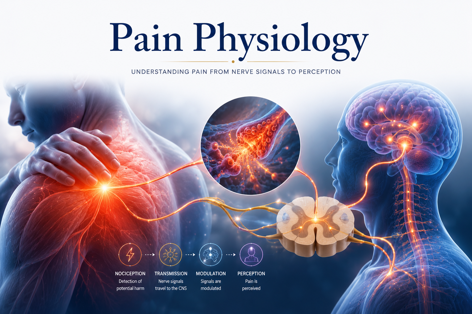
Step 1: Nociception — Detecting Potential Harm
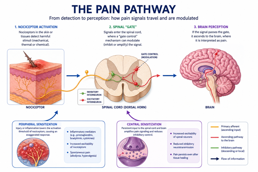
Pain begins with nociception — the detection and transmission of potentially harmful stimuli by specialized sensory neurons called nociceptors.
Nociceptors are free nerve endings found in virtually all tissues of the body — skin, muscle, joints, tendons, ligaments, and viscera. They are activated by:
- Mechanical stimuli: Excessive pressure, stretch, or compression.
- Thermal stimuli: Extreme heat or cold.
- Chemical stimuli: Inflammatory mediators (prostaglandins, bradykinin, substance P, histamine) released by damaged cells, immune cells, and blood vessels.
An important concept is that nociception is not the same as pain. Nociception is a peripheral neural event — the detection of a potentially harmful stimulus. Pain is the brain’s interpretation of that signal. This distinction has profound implications: nociception can occur without pain (such as under general anesthesia), and pain can occur without significant nociception (as in central sensitization).
Once activated, nociceptors send signals along two types of afferent fibers:
- Aδ (A-delta) fibers: Thinly myelinated, fast-conducting (5-30 m/s). Carry the sharp, immediate, well-localized pain of first contact (e.g., the initial stab of touching a sharp object).
- C fibers: Unmyelinated, slow-conducting (0.5-2 m/s). Carry dull, burning, poorly localized pain — the lingering, throbbing pain that follows an injury.
Step 2: Spinal Processing — The Gate
The nociceptive signal travels to the dorsal horn of the spinal cord, where it synapses with second-order neurons. This is not a simple relay station — it is an active processing center where pain signals can be amplified or suppressed before they ascend to the brain.
The gate control theory of pain (Melzack and Wall, 1965) describes one key mechanism: non-painful sensory input (touch, vibration, movement) carried by large-diameter Aβ fibers can “close the gate” on pain signals at the dorsal horn, reducing the nociceptive signal reaching the brain. This is why rubbing an injured area provides temporary relief — and it is one mechanism through which manual therapy, transcutaneous electrical stimulation (TENS), and movement reduce pain.
Descending pain modulation is another critical mechanism. The brain is not a passive recipient of pain signals — it actively modulates them by sending descending signals down the spinal cord through pathways originating in the periaqueductal gray (PAG) and rostroventral medulla (RVM). These descending signals can either inhibit (reduce) or facilitate (amplify) dorsal horn pain transmission. They use endogenous opioids (endorphins and enkephalins) and serotonin/norepinephrine as neurotransmitters.
Exercise — including therapeutic exercise — significantly activates descending pain inhibitory pathways, producing what is called exercise-induced hypoalgesia — a temporary reduction in pain sensitivity that follows exercise. This is one of the powerful pain-relieving mechanisms of physical therapy.
Step 3: Brain Processing — Where Pain Is Created
Pain is ultimately created in the brain through the activation of a distributed network called the pain neuromatrix — including the somatosensory cortex (location and intensity), anterior cingulate cortex (emotional suffering), prefrontal cortex (cognitive appraisal), insula (interoceptive awareness), and amygdala (fear and threat assessment).
Crucially, the brain’s decision to produce pain is based not just on nociceptive input, but on the brain’s assessment of the overall situation — including the perceived threat level, past experiences with pain, emotional state, beliefs about pain, and the social context. Two people with identical tissue injuries can experience dramatically different levels of pain — because their brains interpret the available information differently.
This is not “all in the head” — it is neuroscience. The brain is doing exactly what it is designed to do: using all available information to create the most accurate assessment of threat and generate appropriate behavior. Understanding this helps patients recognize that their pain is real (it always is) but also that it is modifiable through approaches that change the brain’s assessment of threat.
Peripheral Sensitization: The Injured Tissue Becomes More Sensitive
After tissue injury, the inflammatory response lowers the activation threshold of peripheral nociceptors — making them fire more easily and more vigorously. This is called peripheral sensitization and manifests as:
- Allodynia: Pain from stimuli that are normally not painful (e.g., light touch to a sunburned shoulder).
- Primary hyperalgesia: Exaggerated pain response at the site of injury (e.g., extreme sensitivity of the skin around a cut).
Peripheral sensitization is protective — it discourages use of the injured tissue, creating a rest-enforced healing period. Most peripheral sensitization resolves as inflammation resolves and tissue heals.
Central Sensitization: The Nervous System Becomes Amplified
When pain signals are prolonged or intense enough, changes can occur in the spinal cord and brain that amplify pain signals from all areas of the body — not just the original injury site. This is called central sensitization and is a key mechanism in chronic pain conditions.
In central sensitization:
- Spinal cord neurons become hyperexcitable (lower activation threshold, larger receptive fields).
- Inhibitory interneurons lose function (reducing the gate-control dampening of pain).
- Brain processing of pain becomes dysregulated — the pain neuromatrix becomes chronically active even without significant peripheral nociceptive input.
Central sensitization underlies conditions like fibromyalgia, complex regional pain syndrome (CRPS), chronic low back pain, and chronic widespread pain. In these conditions, treatments targeting peripheral tissue (injections, surgery) often fail because the problem is no longer primarily in the tissue — it is in the sensitized nervous system.
How Physical Therapy Targets Pain Mechanisms
Physical therapy is uniquely positioned to address pain at every level:
At the peripheral level: Therapeutic exercise reduces inflammation, promotes tissue healing, and reduces the chemical milieu that drives peripheral sensitization.
At the spinal level: Manual therapy, TENS, and exercise activate Aβ fibers (gate control), stimulate endogenous opioid release, and activate descending inhibitory pathways.
At the brain level: Graded exercise and graded exposure to feared movements progressively reduce the brain’s threat assessment, reducing central sensitization. Pain neuroscience education (PNE) — teaching patients accurate, up-to-date information about pain biology — consistently improves pain, disability, and fear-avoidance in chronic pain populations.
Psychological: Physical therapy that incorporates cognitive-behavioral principles — addressing catastrophizing, fear of movement, and self-efficacy — reduces the psychological amplifiers of pain that operate at the brain level.
Conclusion
Pain is not a simple alarm signal from damaged tissue. It is a sophisticated, adaptive, and modifiable experience created by the brain from a complex interplay of nociceptive signals, emotional state, beliefs, past experience, and social context. Understanding this biology empowers both physical therapists and their patients.
For patients with acute pain, the message is: pain does not mean danger, and gentle movement is safe and healing. For patients with chronic pain, the message is: the nervous system can change, desensitization is possible, and physical therapy — including movement, education, and graded exposure — is among the most effective tools available.
Pain physiology is, ultimately, a story of hope: the same nervous system plasticity that can amplify pain can also be directed, through the right therapeutic approaches, toward its relief.
References
- Hall, J.E., & Hall, M.E. (2020). Guyton and Hall Textbook of Medical Physiology (14th ed.). Elsevier.
- Melzack, R., & Wall, P.D. (1965). Pain mechanisms: A new theory. Science, 150(3699), 971-979.
- Butler, D.S., & Moseley, G.L. (2013). Explain Pain (2nd ed.). Noigroup Publications.
- Costanzo, L.S. (2022). Physiology (7th ed.). Elsevier.
Disclaimer: This article is for educational purposes only and does not constitute medical advice. Always consult a qualified healthcare professional for personal health concerns.
The site is written and curated by Paul Morgan, a graduate in Physiotherapy (2026), with a particular interest in cardiorespiratory and musculoskeletal physical therapy. Every article on this site is grounded in academic physiology and physical therapy coursework. Content reviewed for clinical accuracy before publishing.