Homeostasis: How the Body Maintains Balance During Exercise and Physical Therapy
Learn how homeostasis maintains the body’s internal balance during exercise, injury, and physical therapy — and how therapeutic interventions support these regulatory systems.
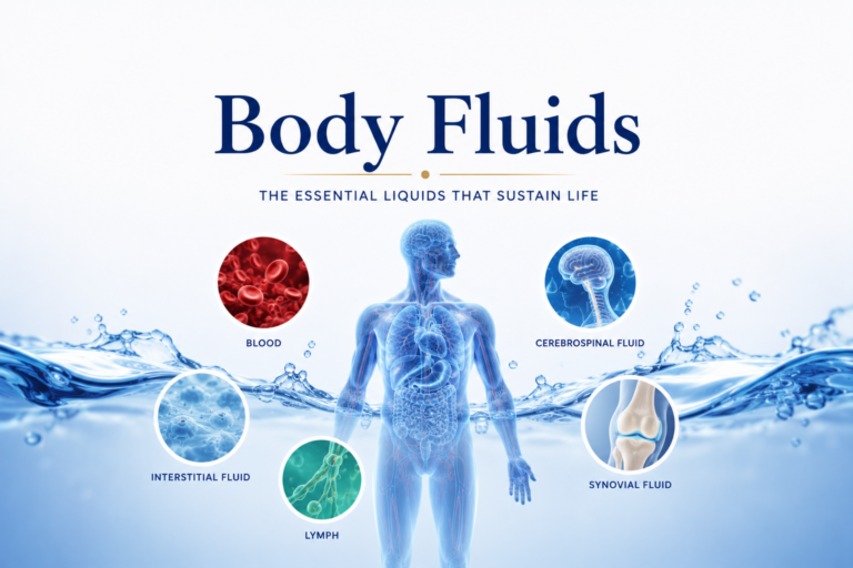
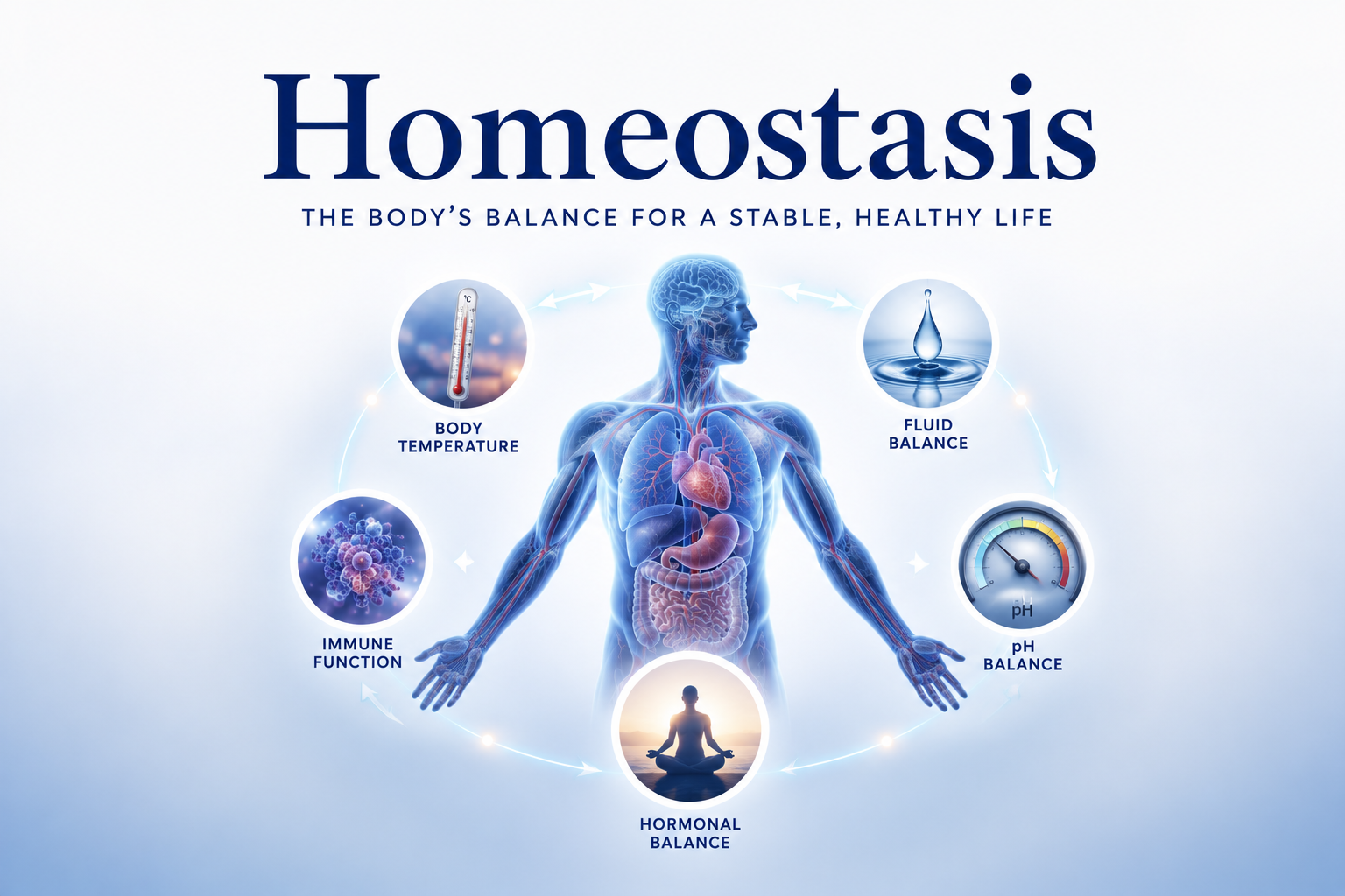
The human body is subject to a constant barrage of internal and external challenges: temperature fluctuations, changing oxygen demands, blood glucose swings, pH shifts from exercise, and the physiological stress of injury. Yet despite this relentless challenge, the body’s internal environment remains remarkably stable. Blood temperature stays within a fraction of a degree. Blood pH fluctuates by only hundredths of a unit. Blood glucose is maintained within a narrow range. This ability to maintain a stable internal environment in the face of changing conditions is called homeostasis, and it is one of the most fundamental properties of living organisms.
Understanding homeostasis is essential for anyone in physical therapy. It explains why the body responds to injury and exercise in predictable ways, how therapeutic interventions interact with the body’s regulatory systems, and why disruptions of homeostasis — as in fever, chronic inflammation, or metabolic disease — can significantly impair rehabilitation.
What Is Homeostasis?
The term homeostasis was coined by physiologist Walter Cannon in 1926, derived from the Greek words for “same” (homeo) and “standing still” (stasis). But it is important to note that homeostasis does not mean absolute constancy — it means dynamic equilibrium. Regulated variables fluctuate within normal ranges and are continuously adjusted in response to internal and external changes.
For example:
- Core body temperature is maintained at approximately 37°C (98.6°F), but can range from ~36.5°C during sleep to ~37.5°C during the afternoon.
- Blood glucose is maintained at approximately 80-120 mg/dL, rising after meals and decreasing between meals, but always returning to this range.
- Blood pH is maintained between 7.35 and 7.45, with momentary fluctuations during intense exercise that are rapidly corrected.
Each of these variables is controlled by homeostatic systems that detect deviations, process information, and implement corrective responses.
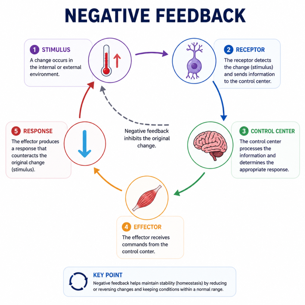
The Components of Homeostatic Control Systems
All homeostatic control systems share three fundamental components:
1. The Sensor (Receptor): A structure that monitors the value of a variable — detecting changes from the set point. Examples include:
- Thermoreceptors in the skin and hypothalamus that detect temperature changes.
- Chemoreceptors in the aortic arch and carotid bodies that detect changes in blood oxygen, CO₂, and pH.
- Baroreceptors in blood vessel walls that detect changes in blood pressure.
- Glucose sensors in pancreatic beta cells that detect blood glucose levels.
2. The Control Center: The structure that receives information from the sensor, compares it to the set point, and determines the appropriate response. In most homeostatic systems, this is the brain (particularly the hypothalamus) or an endocrine organ (like the pancreas). The control center processes afferent signals (incoming from sensors) and generates efferent signals (outgoing to effectors).
3. The Effector: The structure that carries out the corrective response. Effectors include muscles (skeletal and smooth) and glands. Their responses bring the variable back toward the set point.
This system operates through feedback loops — most commonly negative feedback, in which the corrective response counteracts the original deviation, returning the variable to its set point.
Negative Feedback: The Body’s Primary Control Mechanism
Negative feedback is the most common homeostatic mechanism because it provides stability. When a variable deviates from the set point, the response reduces (negates) the deviation and restores equilibrium.
Classic examples relevant to physical therapy:
Temperature regulation: During exercise, body temperature rises due to heat generated by muscle metabolism. Thermoreceptors detect this rise and signal the hypothalamus. The hypothalamus activates effectors: sweat glands (increasing heat loss through evaporative cooling) and cutaneous blood vessels (dilating to increase heat radiation from the skin surface). As body temperature falls back toward normal, these responses diminish.
Physical therapists consider thermoregulation when prescribing exercise in hot environments or for patients with conditions that impair sweating (like some neurological conditions). Exercise-induced hyperthermia can impair performance and, in extreme cases, cause heat illness.
Blood glucose regulation: When blood glucose rises after a meal, pancreatic beta cells detect the increase and secrete insulin. Insulin stimulates muscle and fat cells to take up glucose, and the liver to store glucose as glycogen, returning blood glucose toward normal. When blood glucose falls (during fasting or exercise), glucagon from pancreatic alpha cells mobilizes glycogen and stimulates gluconeogenesis.
For physical therapy patients with diabetes, exercise significantly affects blood glucose regulation. Exercise increases glucose uptake by muscle (both insulin-dependent and independent mechanisms), typically lowering blood glucose. This requires careful management around medication and food intake.
Blood pressure regulation: During exercise, blood pressure rises. Baroreceptors in the aorta and carotid arteries detect the increase and signal the cardiovascular control center in the brainstem. Heart rate and stroke volume are adjusted, and arterioles dilate in active muscles while constricting in less active areas, redistributing blood flow and normalizing pressure.
Positive Feedback: The Exception
While negative feedback restores equilibrium, some homeostatic situations involve positive feedback — where the response amplifies the original deviation rather than reversing it. Positive feedback systems are used in the body when a rapid, self-amplifying process is needed to reach a specific endpoint.
Examples include:
- Blood clotting: Damage activates platelets → platelets release clotting factors → more platelets are activated → clot forms quickly. The process stops when the damaged vessel is sealed.
- Labor contractions: Pressure of the baby’s head → oxytocin release → stronger contractions → more pressure → more oxytocin. Stops after delivery.
Understanding positive feedback helps explain the biology of certain injury responses: the initial inflammatory cascade involves positive feedback elements that amplify the immune response, ensuring rapid, sufficient mobilization of immune cells. Physical therapy interventions that modulate inflammation work partly by interrupting excessive positive feedback amplification.
Homeostasis and the Stress Response
Injury, surgery, and significant illness disrupt homeostasis at multiple levels simultaneously. The body mounts a stress response — coordinated by the hypothalamic-pituitary-adrenal (HPA) axis and the sympathetic nervous system — that adjusts homeostatic set points to meet the extraordinary demands of survival and healing.
This response includes:
- Elevated cortisol: Mobilizes glucose and amino acids for energy, modulates immune function, and reduces certain aspects of inflammation.
- Elevated catecholamines (epinephrine and norepinephrine): Increase heart rate, blood pressure, and glucose availability.
- Altered fluid and electrolyte regulation: The body retains sodium and water (via aldosterone and ADH), contributing to post-injury edema.
Physical therapy interventions — including early mobilization, progressive exercise, and pain education — help the body transition from the acute stress response back to normal homeostatic regulation. Prolonged physiological stress (from chronic pain, psychological distress, or repeated re-injury) can sustain the stress response chronically, impairing healing and rehabilitative progress.
Homeostasis and Exercise: A Dynamic Balance
Exercise is the greatest regular challenge to the body’s homeostatic systems. During vigorous exercise:
- Core temperature rises by 1-2°C
- Blood pH drops from ~7.40 to ~7.35 (and further in intense exercise)
- Blood glucose fluctuates
- Osmolarity increases (from water loss through sweat)
- Lactate rises dramatically
- Oxygen consumption increases 10-20-fold
All of these changes are detected and partially compensated by homeostatic systems — increasing breathing rate, dilating blood vessels, activating sweat glands, mobilizing glucose reserves, and adjusting cardiac output.
Fitness training makes homeostatic responses more efficient. Trained individuals tolerate greater exercise-induced deviations and restore homeostasis more rapidly. This improved regulatory efficiency is one of the mechanisms through which physical therapy progressively improves exercise capacity.
Conclusion
Homeostasis is the physiological foundation on which physical therapy operates. Every exercise session challenges the body’s regulatory systems; every therapeutic intervention either supports or modulates homeostatic mechanisms; and every successful rehabilitation outcome represents the restoration of homeostatic stability in tissues and body systems that had been disrupted by injury or disease.
For physical therapy patients, the practical message is that the body is always working to return to balance — and physical therapy provides the structured stimulus and support that helps it do so more effectively. Understanding homeostasis transforms the body from a mystery into a comprehensible system, one with predictable responses to the right interventions applied at the right time.
References
- Hall, J.E., & Hall, M.E. (2020). Guyton and Hall Textbook of Medical Physiology (14th ed.). Elsevier.
- Costanzo, L.S. (2022). Physiology (7th ed.). Elsevier.
Disclaimer: This article is for educational purposes only and does not constitute medical advice. Always consult a qualified healthcare professional for personal health concerns.
The site is written and curated by Paul Morgan, a graduate in Physiotherapy (2026), with a particular interest in cardiorespiratory and musculoskeletal physical therapy. Every article on this site is grounded in academic physiology and physical therapy coursework. Content reviewed for clinical accuracy before publishing.