Cell Organelles Explained: The Internal Machinery That Drives Healing and Recovery
Discover how cell organelles like the Golgi apparatus, lysosomes, and endoplasmic reticulum work together to support tissue healing and recovery in physical therapy.
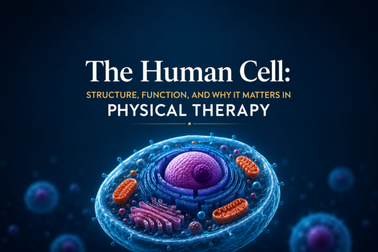
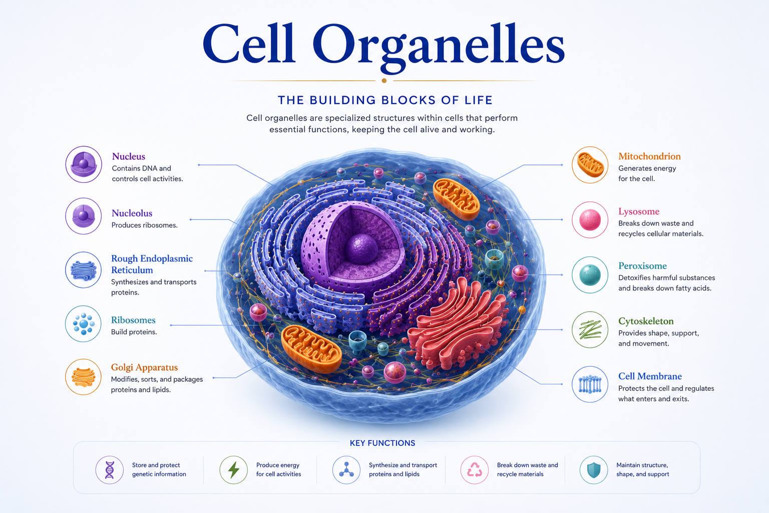
The interior of a human cell is not empty space. It is a bustling, organized metropolis filled with specialized structures — organelles — that each perform a distinct and vital function. Just as a factory floor has separate stations for assembling, packaging, quality control, and waste disposal, the cell has organelles dedicated to specific tasks that collectively keep it alive and allow it to respond to injury, exercise, and healing.
In physical therapy, understanding what these organelles do provides a deeper appreciation of why therapeutic strategies work. This article explores the key organelles of the human cell, their functions, and their relevance to musculoskeletal healing and rehabilitation.
The Endoplasmic Reticulum: The Cell’s Production Network
The endoplasmic reticulum (ER) is an extensive network of membrane-enclosed channels and sacs that winds through the cytoplasm like an interconnected system of tunnels. It is divided into two types with different functions.
Rough endoplasmic reticulum (rough ER) is studded with ribosomes on its outer surface, giving it a bumpy appearance under the microscope. These ribosomes synthesize proteins that are destined for secretion (release outside the cell) or for use in the cell membrane. The rough ER assists in folding these proteins into their correct three-dimensional shapes — a critical step, because misfolded proteins are non-functional and can be toxic to the cell.
In healing tissues, the rough ER is extremely active. Fibroblasts — the cells responsible for producing collagen in tendons, ligaments, and fascia — rely heavily on their rough ER to manufacture and correctly fold the collagen proteins that are essential for structural repair. Increasing collagen production is one of the key goals of physical therapy during the repair and remodeling phases of healing.
Smooth endoplasmic reticulum (smooth ER) lacks ribosomes and has a different set of functions. It synthesizes lipids and phospholipids needed for membrane repair, detoxifies drugs and metabolic waste products, and — critically for muscle function — stores and releases calcium ions. In skeletal muscle cells, the smooth ER is called the sarcoplasmic reticulum. It releases calcium ions in response to nerve signals, triggering muscle contraction. Damage to the sarcoplasmic reticulum — as can happen in severe muscle injuries — impairs calcium handling and leads to prolonged weakness and altered muscle function.
The Golgi Apparatus: The Cell’s Postal Service
The Golgi apparatus (sometimes called the Golgi complex) is a stack of flattened membrane sacs that processes, modifies, and sorts proteins coming from the rough ER. It has been compared to a post office: proteins arrive, receive final modifications (such as the addition of sugar chains in a process called glycosylation), are sorted by destination, and are packaged into vesicles for delivery.
Proteins processed by the Golgi can be directed to three main destinations:
- The cell membrane, where they function as receptors, transport channels, or structural components.
- Lysosomes, where they function as digestive enzymes.
- Outside the cell, through a process called exocytosis — this is how cells secrete hormones, growth factors, and structural proteins like collagen.
In the context of physical therapy, the Golgi apparatus plays a critical role during the remodeling phase of tissue healing. Growth factors and signaling molecules that coordinate cellular repair are secreted via the Golgi pathway. Interventions that promote cellular activity — including therapeutic exercise and some physical therapy modalities — indirectly support this secretory process by maintaining good cellular metabolism and circulation.
Lysosomes: The Cell’s Recycling and Cleanup Crew
Lysosomes are small, spherical vesicles containing powerful digestive enzymes. Their primary function is to break down unwanted materials inside the cell — including damaged organelles, ingested pathogens, and cellular debris.
The process by which lysosomes degrade the cell’s own damaged components is called autophagy (literally “self-eating”). Far from being destructive, autophagy is a critical quality-control mechanism. By removing damaged mitochondria, misfolded proteins, and dysfunctional organelles, autophagy keeps the cell healthy and prevents the buildup of toxic debris.
In physical therapy, lysosomal activity is directly relevant in two scenarios:
After exercise-induced muscle damage, lysosomes in muscle cells become highly active. They clear away damaged protein fragments and cellular debris, preparing the cell for the synthesis of new, functional proteins. This is part of why rest after intense exercise is important — it allows this cellular housekeeping to take place.
In chronic musculoskeletal conditions like tendinopathy, lysosomal dysfunction has been implicated in the failure of normal tissue repair. Research suggests that in chronic tendon disease, normal autophagy is impaired, leading to accumulation of damaged cellular components and perpetuating tissue degeneration. Physical therapy interventions — particularly eccentric exercise — may partly work by stimulating lysosomal and autophagic pathways in tendon cells.
Peroxisomes: Managing Oxidative Stress
Peroxisomes are small organelles that contain enzymes involved in oxidative reactions. One of their most important functions is breaking down hydrogen peroxide (H₂O₂) — a reactive oxygen species that is toxic to cells — into harmless water and oxygen. This is done by an enzyme called catalase, one of the most efficient enzymes known.
Peroxisomes also participate in the metabolism of very long-chain fatty acids and the synthesis of certain lipids. In the liver and kidney, they help detoxify alcohol and other harmful substances.
During intense exercise, the production of reactive oxygen species increases substantially. If the cellular antioxidant systems — including peroxisomes and enzymes like superoxide dismutase — are overwhelmed, oxidative stress occurs, damaging cellular membranes, proteins, and DNA. Physical therapy programs that incorporate appropriate exercise intensity and progression help the body’s antioxidant capacity adapt over time, reducing oxidative stress and supporting cellular health.
Ribosomes: The Cell’s Protein Factories
Ribosomes are not technically membrane-bound organelles, but they are among the most important structures in the cell. They are the molecular machines that translate genetic instructions from mRNA into protein chains.
Ribosomes can be found either free in the cytoplasm (producing proteins used within the cell) or attached to the rough ER (producing proteins destined for secretion or the cell membrane). During periods of active repair and growth, cells upregulate ribosome production to increase their protein-synthesizing capacity.
In skeletal muscle, ribosome content is closely linked to muscle growth capacity. After a period of disuse — such as immobilization following surgery — muscle cells lose ribosomes along with muscle mass. Resistance exercise during rehabilitation stimulates ribosomal biogenesis (the production of new ribosomes), which is an early step in the restoration of muscle protein synthesis capacity.
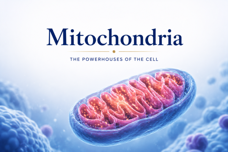
Mitochondria and Peroxisomes: Partners in Energy and Cleanup
Mitochondria and peroxisomes work together in fatty acid metabolism. Very long-chain fatty acids are first partially processed in peroxisomes and then completed in mitochondria through beta-oxidation. This coordination ensures efficient energy extraction from fats — important during prolonged, low-intensity aerobic exercise, which is often prescribed in physical therapy to promote recovery and cardiovascular fitness.
Conclusion
The internal organelles of the cell function as an integrated team. The rough ER produces proteins, the Golgi processes and ships them, lysosomes clear away the debris, peroxisomes neutralize oxidative stress, ribosomes synthesize the molecular tools for repair, and mitochondria power the entire operation. Together, these organelles create the cellular environment in which healing happens.
Physical therapy is, at its core, a science of stimulating and supporting these cellular processes through strategic mechanical and physiological interventions. When a therapist prescribes progressive loading, manual therapy, or aerobic conditioning, the effects cascade through every level of biology — including the organelles described in this article. The more we understand these processes, the more precisely and effectively we can support recovery.
Disclaimer: This article is for educational purposes only and does not constitute medical advice. Always consult a qualified healthcare professional for personal health concerns.