The Plasma Membrane: How Cells Communicate and What It Means for Healing
Discover how the plasma membrane controls what enters and exits cells, and how this boundary plays a key role in inflammation, healing, and physical therapy.
Imagine a city surrounded by a sophisticated wall — a barrier that controls who enters, who exits, and what messages pass through its gates. That is essentially what the plasma membrane does for every cell in your body. This thin but extraordinarily complex structure is the cell’s first line of defense, its communication hub, and its primary gateway to the outside world.
In the context of physical therapy and musculoskeletal health, the plasma membrane plays a central role in processes like inflammation, pain signaling, nutrient uptake, and tissue repair. Understanding how it works helps explain many of the physiological effects that physical therapy treatments produce.
Structure of the Plasma Membrane
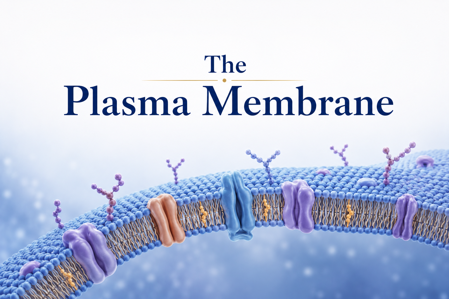
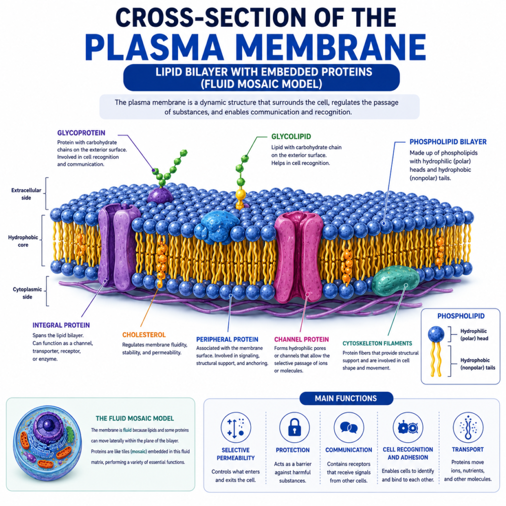
The plasma membrane is made of a phospholipid bilayer — two layers of phospholipid molecules arranged tail-to-tail. Each phospholipid has a hydrophilic (water-attracting) head and a hydrophobic (water-repelling) tail. This arrangement creates a stable barrier that separates the cell’s interior from the outside environment.
Embedded within this lipid bilayer are various proteins that give the membrane its remarkable functionality. Some proteins act as channels that allow specific molecules to pass through. Others function as receptors that detect signals from hormones, neurotransmitters, or growth factors. Still others serve as enzymes, identification markers (like blood type antigens), or structural anchors that connect the membrane to the cell’s internal skeleton.
This model — known as the fluid mosaic model — describes the membrane as a dynamic, constantly shifting structure rather than a rigid wall. The fluidity of the membrane is essential: it allows proteins to move laterally, membranes to fuse together, and the cell to respond quickly to changing conditions.
How the Membrane Controls What Enters and Exits
One of the most critical functions of the plasma membrane is selective permeability — the ability to control which substances can cross it. This is vital for maintaining the internal environment of the cell and, by extension, the health of tissues throughout the body.
Small, nonpolar molecules like oxygen and carbon dioxide can cross the membrane freely through a process called simple diffusion. This is how muscle cells receive oxygen during aerobic exercise and release CO2 as a byproduct.
Larger or charged molecules — like glucose, amino acids, and ions such as sodium, potassium, and calcium — need help crossing the membrane. They pass through specialized protein channels or carriers via a process called facilitated diffusion or, in some cases, active transport (which requires energy in the form of ATP).
In physical therapy, this matters enormously. When tissues are compressed, stretched, or stimulated through exercise, the movement of ions and nutrients across cell membranes changes. For example, calcium ions flowing into muscle cells through membrane channels trigger muscle contraction. Disrupting this flow — as happens in some muscle injuries — leads to impaired contraction and pain.
The Membrane and Inflammation
When tissues are injured, the plasma membrane becomes a central actor in the inflammatory response. Physical trauma ruptures cell membranes, releasing their contents into the surrounding tissue. This triggers the release of signaling molecules — including prostaglandins and cytokines — that attract immune cells to the site and initiate the healing process.
The membrane is also where many anti-inflammatory drugs act. Nonsteroidal anti-inflammatory drugs (NSAIDs), for instance, block enzymes (like COX-1 and COX-2) that are associated with the membrane and involved in prostaglandin synthesis. Understanding this mechanism helps explain why physical therapists often coordinate with physicians regarding medication timing and therapeutic exercise.
Additionally, membrane receptors play a key role in pain signaling. Nociceptors — the sensory receptors that detect harmful stimuli — are located on the membranes of specialized neurons. When these receptors are activated by mechanical pressure, heat, or chemical irritants released during injury, they send pain signals to the brain. Physical therapy interventions like manual therapy, mobilization, and therapeutic exercise can modulate these receptor activities, contributing to pain relief.
Membrane Permeability and Edema
Edema — the accumulation of excess fluid in tissues — is a common consequence of injury and one of the main challenges addressed in physical therapy. One of its key causes is increased membrane permeability.
When cells are damaged or when the immune system releases specific molecules, the membranes of endothelial cells (which line blood vessels) become more permeable. This allows fluid from the blood to leak into surrounding tissue, causing swelling. Increased membrane permeability also occurs in muscle cells after intense exercise, contributing to delayed onset muscle soreness (DOMS).
Physical therapy treatments such as compression, elevation, lymphatic drainage massage, and early controlled movement all work, in part, by helping restore normal membrane permeability and facilitating the movement of excess fluid back into the lymphatic and venous systems.
Mechanotransduction: How Membranes Sense Movement
One of the most exciting areas of research in physical therapy involves mechanotransduction — the process by which mechanical forces (like those applied during exercise or manual therapy) are converted into biochemical signals at the cellular level.
The plasma membrane plays a central role in this process. Specialized proteins called integrins span the membrane and connect the cell’s internal skeleton (the cytoskeleton) to the extracellular matrix — the network of proteins that surrounds cells. When the extracellular matrix is deformed by mechanical loading (such as during exercise or soft tissue manipulation), integrins transmit these forces across the membrane into the cell, activating signaling pathways that regulate gene expression, protein synthesis, and cell behavior.
This is one reason why therapeutic exercise is so effective in tissue repair: the mechanical forces generated by movement activate cellular responses that accelerate healing. The plasma membrane is the sensor that detects these forces and translates them into biological instructions.
Cell Communication Through the Membrane
Cells constantly communicate with each other through the plasma membrane. Receptor proteins on the membrane surface detect chemical signals — including hormones like cortisol and adrenaline, growth factors like IGF-1, and neurotransmitters like acetylcholine — and translate them into changes in cell behavior.
In rehabilitation, this communication system is essential. Growth factors released after injury bind to membrane receptors on fibroblasts (connective tissue cells) and muscle cells, stimulating them to divide, migrate to the injury site, and produce new tissue. Therapeutic strategies that promote blood flow — including exercise, heat therapy, and ultrasound — enhance the delivery of these signaling molecules to injured tissues.
Conclusion
The plasma membrane is far more than a simple boundary around the cell. It is a sophisticated, dynamic system that controls the cell’s interactions with the world around it — regulating what enters and exits, sensing mechanical forces, transmitting pain signals, coordinating the inflammatory response, and facilitating tissue repair.
For physical therapists and their patients, understanding the plasma membrane provides insight into why therapeutic interventions work at a biological level. From the way exercise triggers healing signals to the way manual therapy reduces pain, the plasma membrane is always part of the story.
Taking care of your body through movement, proper hydration, and appropriate physical therapy is, ultimately, taking care of your cells — one membrane at a time.
References
- Alberts, B., Heald, R., Johnson, A., Morgan, D., Raff, M., Roberts, K., & Walter, P. (2022). Molecular Biology of the Cell (7th ed.). W.W. Norton & Company.
- Ross, M.H., & Pawlina, W. (2020). Histology: A Text and Atlas (8th ed.). Wolters Kluwer.
Disclaimer: This article is for educational purposes only and does not constitute medical advice. Always consult a qualified healthcare professional for personal health concerns.
The site is written and curated by Paul Morgan, a graduate in Physiotherapy (2026), with a particular interest in cardiorespiratory and musculoskeletal physical therapy. Every article on this site is grounded in academic physiology and physical therapy coursework. Content reviewed for clinical accuracy before publishing.