The Human Cell: Structure, Function, and Why It Matters in Physical Therapy
Learn how the human cell works, its main components, and why understanding cell biology is essential for physical therapy and rehabilitation.
Every living thing on Earth is made of cells. The human body contains approximately 37 trillion of them. But what exactly is a cell, and why should someone interested in physical therapy care about it? The answer is simple: every injury, every recovery, every therapeutic exercise you perform ultimately happens at the cellular level. Understanding the basic unit of life gives us a deeper appreciation of how the body heals and how physical therapy works.
In this article, we will break down the structure and function of the human cell, explain the key components that keep it running, and connect that knowledge to what happens during injury and rehabilitation.
What Is a Cell?
A cell is the smallest structural and functional unit of a living organism. It is the basic building block of all tissues, organs, and body systems. Cell biology — the science that studies cells — tells us that all cells share common features, even though they look and behave very differently depending on where they are in the body.
In the human body, cells are classified as eukaryotic cells. This means they have a well-defined nucleus enclosed by a membrane, which contains the genetic information that controls everything the cell does. Compared to prokaryotic cells (like bacteria), eukaryotic cells are more complex, larger, and have specialized compartments called organelles that carry out specific functions.
Understanding this distinction matters in physiotherapy because certain infections — caused by bacteria or viruses — can disrupt normal cellular function, slow down healing, and complicate recovery.
The Three Main Parts of a Human Cell
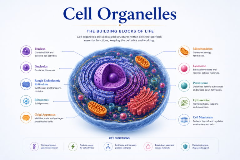
Every human cell can be divided into three main regions: the plasma membrane, the cytoplasm, and the nucleus.
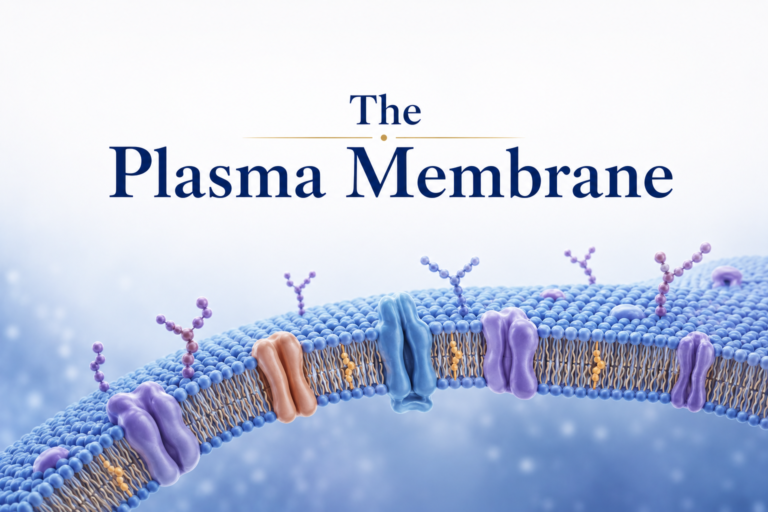
The plasma membrane is the outer boundary of the cell. It is made of a double layer of lipid molecules with proteins embedded in it — a structure known as the fluid mosaic model. This membrane is selectively permeable, meaning it controls what enters and exits the cell. In physical therapy, this is relevant because nutrients, oxygen, and therapeutic molecules must cross this barrier to support healing. When the membrane is damaged — as happens in muscle injuries — the cell loses its ability to regulate its internal environment.
The cytoplasm is the gel-like substance that fills the cell and contains all the organelles. It is where most of the cell’s metabolic activities take place. The cytoplasm also acts as a transport medium, moving molecules from one organelle to another.
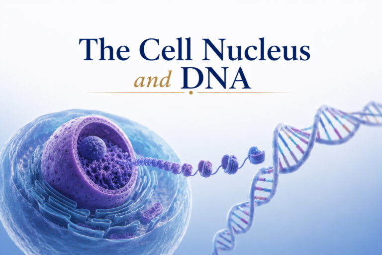
The nucleus is the control center of the cell. It houses the cell’s DNA — the genetic blueprint that instructs the cell on how to build proteins, respond to injury, divide, and repair itself. In the context of rehabilitation, the nucleus plays a crucial role in activating genes that promote tissue healing and muscle adaptation.
Key Organelles and Their Functions
Inside the cytoplasm, several organelles work together to keep the cell alive and functional. Here are the most important ones for understanding physical therapy:
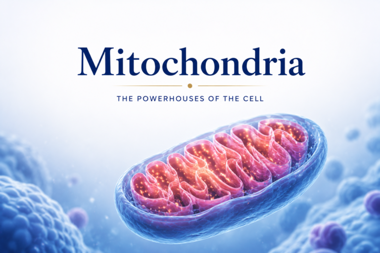
Mitochondria are often called the powerhouses of the cell. They produce adenosine triphosphate (ATP), the energy molecule that fuels every cellular process — including muscle contraction. During physical therapy exercises, the demand for ATP increases significantly. Muscles with more mitochondria are more resistant to fatigue and recover faster.
Ribosomes are tiny structures that build proteins based on instructions from the nucleus. Proteins are essential for muscle repair, enzyme production, and tissue regeneration. After a muscle injury, ribosomes work overtime to produce the proteins needed for healing.
The endoplasmic reticulum (ER) comes in two types: rough ER (which is covered in ribosomes and helps produce proteins) and smooth ER (which synthesizes lipids and detoxifies harmful substances). In muscle cells, the smooth ER — known as the sarcoplasmic reticulum — is especially important because it stores and releases calcium, which is essential for muscle contraction.
The Golgi apparatus processes, packages, and ships proteins produced by the ER to their destinations inside or outside the cell. In healing tissues, the Golgi is very active as it prepares and exports proteins needed to rebuild damaged structures.
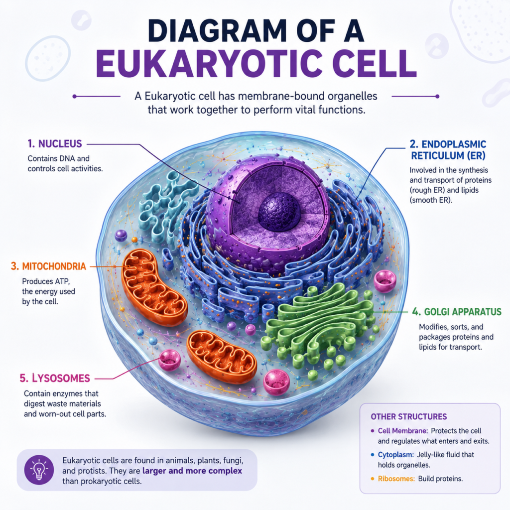
Lysosomes are the cell’s recycling centers. They contain enzymes that break down damaged organelles and debris. After a muscle injury, lysosomes help clear away damaged cellular material so that repair can begin — a process that physical therapy supports through movement and controlled loading.
Cell Types Relevant to Physical Therapy
Not all cells are the same. Some are especially relevant to physical therapy practice:
Muscle cells (myocytes) are long, cylindrical cells packed with proteins that allow contraction. They are among the most metabolically active cells in the body.
Fibroblasts are connective tissue cells responsible for producing collagen. They are critical in the repair of tendons, ligaments, and fascia — tissues frequently treated in physical therapy.
Neurons transmit electrical signals and are involved in pain perception, motor control, and sensory function. Many physical therapy interventions, including nerve mobilization and neuromuscular re-education, directly target neuronal function.
Osteoblasts and osteoclasts are bone cells that build and break down bone tissue, respectively. Understanding their activity is essential when treating fractures or osteoporosis.
Why Cell Health Matters for Recovery
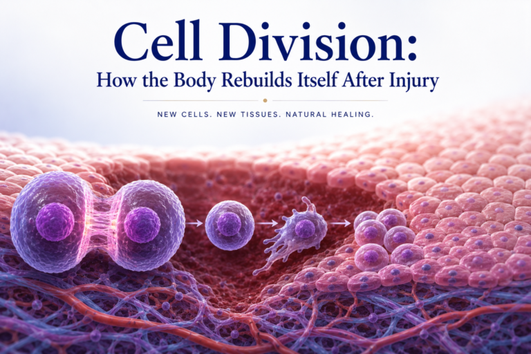
When a tissue is injured, the damage always begins at the cellular level. Cells are disrupted, membranes are torn, organelles malfunction, and the normal flow of cellular activity is interrupted. The body’s healing response — including inflammation, tissue repair, and remodeling — is coordinated by signals sent between cells.
Physical therapy interventions work, in part, by influencing cellular processes. Therapeutic exercise stimulates mitochondrial biogenesis (the creation of new mitochondria), increases protein synthesis in muscle cells, enhances blood flow that delivers oxygen and nutrients to healing cells, and activates mechanoreceptors that send signals to the nervous system.
In other words, when a physical therapist prescribes exercise or manual therapy, the effects go all the way down to the cellular level. Movement is medicine — and the cell is where that medicine is received.
Conclusion
The human cell is far more than a tiny dot under a microscope. It is a complex, dynamic, self-regulating system that underpins every function in the human body. From the energy-producing mitochondria to the protein-building ribosomes, every organelle plays a role in keeping us healthy and helping us recover from injury.
For anyone involved in physical therapy — whether as a practitioner or a patient — understanding cell biology provides a powerful framework for understanding why therapeutic strategies work. When you exercise during rehabilitation, you are not just moving muscles. You are sending signals deep into the cellular machinery that drives your recovery.
References
- Alberts, B., Heald, R., Johnson, A., Morgan, D., Raff, M., Roberts, K., & Walter, P. (2022). Molecular Biology of the Cell (7th ed.). W.W. Norton & Company.
- Ross, M.H., & Pawlina, W. (2020). Histology: A Text and Atlas (8th ed.). Wolters Kluwer.
Disclaimer: This article is for educational purposes only and does not constitute medical advice. Always consult a qualified healthcare professional for personal health concerns.
The site is written and curated by Paul Morgan, a graduate in Physiotherapy (2026), with a particular interest in cardiorespiratory and musculoskeletal physical therapy. Every article on this site is grounded in academic physiology and physical therapy coursework. Content reviewed for clinical accuracy before publishing.