Cell Division: How the Body Rebuilds Itself After Injury
Learn how cell division enables the body to repair damaged tissues, replace worn-out cells, and rebuild after injury — and how physical therapy supports this process.
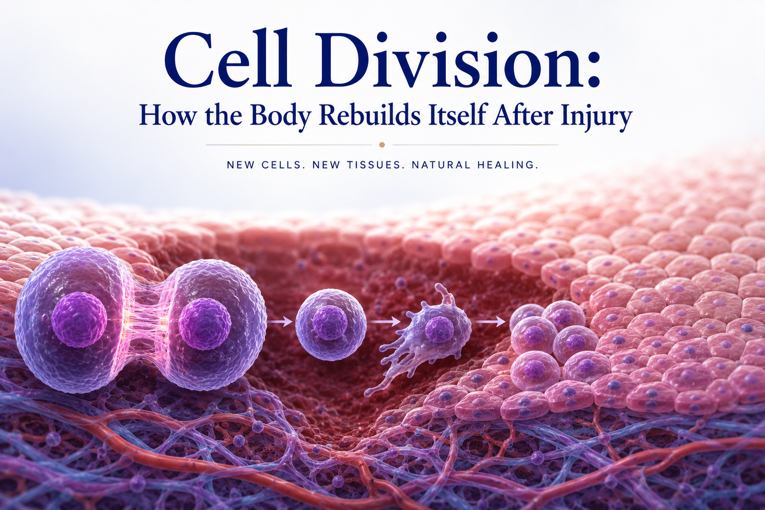
Every time you cut your finger, strain a muscle, or fracture a bone, your body launches a remarkable repair operation. Damaged cells are replaced, new tissue is built, and the injured structure gradually returns to function. At the heart of this process is cell division — the mechanism by which cells reproduce themselves to generate new cells for growth, maintenance, and repair.
Understanding cell division is not merely an academic exercise. For anyone undergoing physical therapy or recovering from injury, it provides a framework for understanding the biological timeline of healing, why certain interventions accelerate recovery, and why pushing too hard too soon can be counterproductive at the cellular level.
The Cell Cycle: Life from a Cell’s Perspective
All cells go through a life cycle called the cell cycle. This cycle consists of two main phases: interphase (the preparatory period) and the mitotic phase (when division actually occurs).
Interphase occupies most of the cell’s life. It consists of three sub-phases:
- G1 phase (Gap 1): The cell grows in size and carries out its normal functions. It also begins preparing for DNA replication.
- S phase (Synthesis): The cell replicates its entire DNA, producing an exact copy of all 46 chromosomes.
- G2 phase (Gap 2): The cell continues to grow and checks that DNA replication was completed accurately before proceeding to division.
After interphase, the cell enters the mitotic phase, in which it divides into two genetically identical daughter cells. This division involves two processes: mitosis (division of the nucleus) and cytokinesis (division of the cytoplasm).
The entire cycle takes anywhere from 12 to 24 hours in actively dividing cells, though some specialized cells — like neurons — normally do not divide at all in adults, while others — like intestinal epithelial cells — divide constantly.
Mitosis: The Mechanics of Cell Division
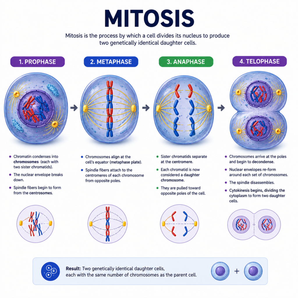
Mitosis is divided into several distinct stages: prophase, metaphase, anaphase, telophase, and cytokinesis. Together, these stages ensure that each daughter cell receives an accurate, complete copy of the genetic information.
During prophase, the duplicated chromosomes condense and become visible under a microscope. The nuclear envelope breaks down, and the mitotic spindle — a structure made of protein fibers — begins to form.
During metaphase, chromosomes align along the center of the cell. This alignment is checked by cellular quality control mechanisms that ensure each chromosome is properly attached to spindle fibers before division proceeds.
During anaphase, the spindle fibers contract and pull the duplicated chromosomes apart to opposite ends of the cell.
During telophase and cytokinesis, the nuclear envelope reforms around each set of chromosomes, the chromosomes decondense, and the cell physically splits into two new cells, each with a complete set of DNA.
Errors during this process — such as chromosomes being distributed unequally — can lead to cell death or, in some cases, cancerous transformation. The cell has multiple checkpoints throughout the cycle to detect and correct errors before they propagate.
Cell Division in Tissue Repair
After an injury, tissue repair depends critically on the ability of resident cells to divide and produce new cells to replace those that were lost or damaged. Different tissues have different capacities for regeneration based on their cellular division ability.
High-regenerative tissues — including skin, bone, and skeletal muscle (through satellite cell activation) — contain populations of stem cells or progenitor cells that can divide extensively in response to injury. Physical therapy is most commonly applied to these tissues and can significantly influence the pace and quality of their repair.
Low-regenerative tissues — such as cartilage and peripheral nerve tissue — have more limited division capacity. Cartilage has very few cells (chondrocytes), which divide slowly and are poorly supplied with blood. This explains why cartilage injuries heal so slowly and why physical therapy for cartilage conditions focuses on preserving function and managing symptoms rather than rapidly restoring tissue.
Non-regenerative tissues — such as the central nervous system — contain neurons that do not divide in adults. Recovery from neurological injuries relies on other mechanisms, such as synaptic plasticity and reorganization of neural circuits, which physical therapy supports through repetitive, task-specific exercises.
The Role of Growth Factors in Stimulating Division
Cell division does not happen spontaneously — it requires specific signals from the cellular environment. These signals come in the form of growth factors: proteins secreted by neighboring cells, immune cells, and platelets that bind to receptors on the target cell’s surface and activate the cell cycle.
Key growth factors in musculoskeletal healing include:
- Platelet-derived growth factor (PDGF): Released by platelets during the initial inflammatory phase, PDGF stimulates fibroblast and smooth muscle cell division.
- Fibroblast growth factor (FGF): Promotes the division of fibroblasts and endothelial cells, essential for the formation of new blood vessels (angiogenesis) in healing tissue.
- Transforming growth factor beta (TGF-β): Stimulates collagen production and modulates the inflammatory response.
- Insulin-like growth factor 1 (IGF-1): Promotes muscle cell division and protein synthesis, and is significantly upregulated during resistance exercise.
Physical therapy interventions influence the production and distribution of these growth factors. Exercise increases local blood flow, bringing more growth factors to healing tissues. Mechanical loading stimulates cells to produce and release their own growth factors. This is one of the key reasons why appropriate early mobilization and therapeutic exercise accelerate healing.
When Cell Division Goes Wrong: Relevance to Physical Therapy
Understanding cell division also helps explain some of the complications that can arise during healing. When the balance between cell division and cell death is disrupted, abnormal tissue responses can occur.
Excessive scar tissue (fibrosis) results from overactive fibroblast division and collagen production. This is common in poorly managed soft tissue injuries and can lead to restricted range of motion and chronic pain. Physical therapy techniques such as soft tissue mobilization, stretching, and progressive loading help remodel scar tissue and prevent excessive fibrosis.
Tendinopathy involves an abnormal proliferation of cells and disorganized collagen production in tendon tissue. Rather than normal healing, the tendon undergoes a degenerative response. Physical therapy — particularly eccentric exercise — has been shown to help normalize cellular activity and improve tendon structure in these cases.
Stress fracture healing requires a carefully regulated sequence of bone cell division (osteoblasts building new bone while osteoclasts remove damaged bone). Too much loading before sufficient cell division has restored bone density can cause re-fracture, while too little loading can delay the hormonal and mechanical signals that stimulate osteoblast activity.
Supporting Cell Division Through Recovery Practices
Physical therapy supports healthy cell division through several mechanisms:
- Progressive loading provides the mechanical signals that stimulate growth factor release and activate the cell cycle in muscle, tendon, and bone cells.
- Blood flow promotion (through exercise, heat, and manual therapy) ensures that the oxygen, nutrients, and signaling molecules needed for cell division reach the healing tissue.
- Adequate rest and sleep provide the time during which DNA replication and cell division occur. Many cells carry out division preferentially during sleep.
- Nutrition — particularly adequate protein, vitamin C (for collagen synthesis), vitamin D (for bone cells), and zinc — supports the cellular machinery needed for division and repair.
Conclusion
Cell division is the biological engine of tissue repair. Without it, injuries would not heal, muscles would not grow, and the body would have no way to replace worn-out or damaged cells. Understanding the cell cycle and how it is regulated helps explain the timeline of recovery, the importance of appropriate loading and rest, and the cellular mechanisms by which physical therapy accelerates healing.
Every therapeutic exercise prescribed in a rehabilitation program, every session of manual therapy, every moment of guided movement — all of these interventions ultimately work by providing the cellular environment and mechanical stimuli that support healthy cell division and tissue regeneration.
References
- Alberts, B., Heald, R., Johnson, A., Morgan, D., Raff, M., Roberts, K., & Walter, P. (2022). Molecular Biology of the Cell (7th ed.). W.W. Norton & Company.
- Ross, M.H., & Pawlina, W. (2020). Histology: A Text and Atlas (8th ed.). Wolters Kluwer.
Disclaimer: This article is for educational purposes only and does not constitute medical advice. Always consult a qualified healthcare professional for personal health concerns.
The site is written and curated by Paul Morgan, a graduate in Physiotherapy (2026), with a particular interest in cardiorespiratory and musculoskeletal physical therapy. Every article on this site is grounded in academic physiology and physical therapy coursework. Content reviewed for clinical accuracy before publishing.