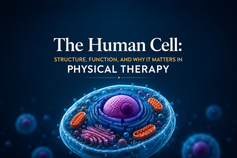
Mitochondria: The Energy Factories Behind Muscle Performance and Recovery
Explore how mitochondria produce energy for muscles, why they matter in physical therapy, and how exercise and rehabilitation influence mitochondrial function.
Ask anyone what mitochondria do, and you will likely hear the same answer: “They are the powerhouse of the cell.” But this famous phrase barely scratches the surface. Mitochondria are extraordinary organelles — dynamic, self-reproducing structures that not only generate the energy that powers every movement you make, but also regulate cell survival, control the inflammatory response, and play a central role in how muscles adapt to exercise and recover from injury.
For anyone engaged in physical therapy — as a practitioner or a patient — understanding mitochondria is essential. These tiny structures explain why muscles fatigue, why aerobic fitness accelerates recovery, and why therapeutic exercise is one of the most powerful tools available to the human body.
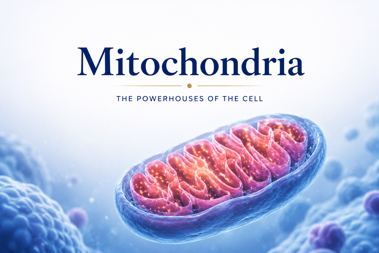
What Are Mitochondria?
Mitochondria are membrane-bound organelles found in nearly all eukaryotic cells. They are particularly abundant in cells with high energy demands — most notably, in muscle cells and neurons. A typical muscle cell may contain hundreds to thousands of mitochondria, while cells with lower energy needs have far fewer.
What makes mitochondria uniquely interesting from a biological standpoint is that they have their own DNA. This is strong evidence supporting the endosymbiotic theory, which proposes that mitochondria were once free-living bacteria that were incorporated into larger cells billions of years ago — a partnership that proved so beneficial it was maintained throughout evolution.
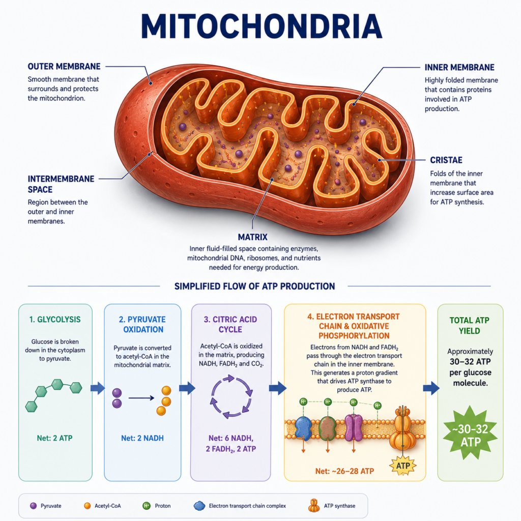
Mitochondria are enclosed by two membranes: a smooth outer membrane and a highly folded inner membrane. The folds of the inner membrane are called cristae, and they significantly increase the surface area available for energy production. Inside the inner membrane is the mitochondrial matrix, where many of the chemical reactions that generate energy take place.
How Mitochondria Produce Energy
The primary function of mitochondria is to produce adenosine triphosphate (ATP) — the molecule that cells use to power virtually every biological process. This energy production occurs mainly through a process called oxidative phosphorylation, which takes place on the inner mitochondrial membrane.
The process begins with nutrients — primarily glucose and fatty acids — being broken down through metabolic pathways (glycolysis in the cytoplasm, and the Krebs cycle in the mitochondrial matrix). These processes generate electron-carrying molecules (NADH and FADH2) that feed into the electron transport chain located on the inner membrane. As electrons pass along this chain, they drive the pumping of protons across the membrane, creating an electrochemical gradient. The energy stored in this gradient is used by an enzyme called ATP synthase to produce ATP.
This process is extraordinarily efficient. The complete oxidation of one glucose molecule through aerobic respiration can yield up to 30-32 molecules of ATP. Compare this to the 2 ATP molecules produced during anaerobic glycolysis (without oxygen), and you can see why aerobic metabolism — which relies on mitochondria — is so much more sustainable for prolonged physical activity.
Mitochondria and Muscle Fatigue
Muscle fatigue during exercise is a complex phenomenon with multiple causes. Mitochondria are at the heart of several of them. When exercise intensity exceeds the capacity of the aerobic system to produce ATP, the body shifts toward anaerobic pathways. This produces energy more quickly but generates lactate and hydrogen ions as byproducts, lowering muscle pH and impairing contractile function — contributing to the burning sensation familiar to anyone who has pushed through a hard workout.
Importantly, the mitochondria themselves can also become damaged during very intense or prolonged exercise. Reactive oxygen species (ROS) — byproducts of high-rate energy production — can damage mitochondrial membranes and DNA. This is one of the cellular mechanisms behind exercise-induced muscle damage and delayed onset muscle soreness (DOMS).
Physical therapy plays a direct role here. Controlled therapeutic exercise, progressive loading, and adequate recovery time all help balance mitochondrial stress with adaptation. This leads to the training effect: the mitochondria become more numerous and more efficient over time.
Mitochondrial Adaptations to Exercise
One of the most remarkable aspects of mitochondria is their plasticity — their ability to change in response to physiological demands. Regular aerobic exercise triggers a process called mitochondrial biogenesis: the production of new mitochondria within muscle cells.
This process is driven by a protein called PGC-1α (peroxisome proliferator-activated receptor gamma coactivator 1-alpha), which is activated during exercise and stimulates the expression of genes involved in mitochondrial production. The result is that trained individuals have more mitochondria per muscle cell, a greater capacity for aerobic energy production, and significantly improved resistance to fatigue.
In physical therapy, this adaptation is one of the key goals of aerobic conditioning programs. Patients recovering from surgery, prolonged immobilization, or serious injury often experience significant mitochondrial loss in affected muscles — a condition known as mitochondrial depletion. Carefully prescribed exercise is the most effective intervention to reverse this and restore normal mitochondrial density.
Mitochondria and Tissue Healing
Beyond energy production, mitochondria play important roles in regulating cell survival and death — processes that are critical during tissue healing. When cells are severely damaged, mitochondria can trigger apoptosis (programmed cell death), which clears away irreparably damaged cells and creates space for regeneration. This controlled process is essential for normal tissue repair.
Mitochondria also influence the inflammatory response. Damaged mitochondria release signals that can amplify inflammation, while healthy mitochondria help resolve it. This is one reason why maintaining good aerobic fitness — which promotes mitochondrial health — is associated with more efficient recovery from musculoskeletal injuries.
Physical therapy modalities such as low-level laser therapy (LLLT) and photobiomodulation have been shown in research to directly stimulate mitochondrial function, increasing ATP production in injured tissues and accelerating the healing process.
Practical Implications for Physical Therapy Patients
Understanding mitochondria has practical implications for anyone undergoing physical rehabilitation:
Aerobic exercise matters. Even patients recovering from orthopedic injuries benefit from maintaining cardiovascular fitness during recovery. Activities like swimming, cycling, or arm ergometry preserve mitochondrial density in unaffected muscles and support systemic recovery.
Progressive overload drives adaptation. Gradually increasing the intensity and duration of exercise is the key to stimulating mitochondrial biogenesis. Physical therapists prescribe this progression carefully to maximize adaptation while avoiding re-injury.
Nutrition supports mitochondrial function. Mitochondria require specific nutrients — including B vitamins, iron, magnesium, and antioxidants — to function optimally. Good nutrition during rehabilitation supports the cellular energy production needed for healing.
Sleep is essential. Much of the cellular repair that follows physical therapy occurs during sleep, when energy demands are lower and mitochondrial repair pathways are most active.
Conclusion
Mitochondria are far more than just the powerhouses of the cell. They are dynamic, adaptable organelles that govern energy production, regulate cell survival, influence inflammation, and drive the physiological adaptations that make physical therapy so effective.
Every time you complete a therapeutic exercise, you are sending signals that reach deep into the mitochondrial machinery of your muscle cells. Over time, these signals build a more efficient, fatigue-resistant, and resilient musculoskeletal system. Understanding this process empowers both therapists and patients to approach rehabilitation with a clearer sense of what is actually happening at the cellular level — and why every session counts.
References
- Alberts, B., Heald, R., Johnson, A., Morgan, D., Raff, M., Roberts, K., & Walter, P. (2022). Molecular Biology of the Cell (7th ed.). W.W. Norton & Company.
- Ross, M.H., & Pawlina, W. (2020). Histology: A Text and Atlas (8th ed.). Wolters Kluwer.
Disclaimer: This article is for educational purposes only and does not constitute medical advice. Always consult a qualified healthcare professional for personal health concerns.
The site is written and curated by Paul Morgan, a graduate in Physiotherapy (2026), with a particular interest in cardiorespiratory and musculoskeletal physical therapy. Every article on this site is grounded in academic physiology and physical therapy coursework. Content reviewed for clinical accuracy before publishing.